Pancreatic Cyst
Pancreatic cysts are increasingly detected with modern imaging. While most are benign, some carry malignant potential. Specialized evaluation and appropriate surveillance are essential for safe, evidence-based management.
Book a Consultation
Understanding Pancreatic Cysts
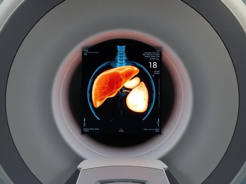
Pancreatic cysts are fluid-filled sacs within the pancreas. They are found in approximately 2-3% of abdominal imaging studies and become more common with age. The key challenge is distinguishing benign cysts from those with malignant potential, as some can progress to pancreatic cancer if left untreated.
Accurate characterisation requires a combination of imaging, cyst fluid analysis, and clinical assessment. Dr. Prem provides comprehensive evaluation using the latest international guidelines to determine appropriate surveillance intervals or need for intervention.
Types of Pancreatic Cysts
Intraductal Papillary Mucinous Neoplasm (IPMN)
Risk: Moderate to HighThe most common type, arising from the pancreatic duct. Can be main-duct, branch-duct, or mixed type with varying malignant potential.
Mucinous Cystic Neoplasm (MCN)
Risk: Moderate to HighOccurs predominantly in women, typically in the body or tail of the pancreas. Has malignant potential and often requires surgical resection.
Serous Cystadenoma
Risk: Very LowBenign tumours with very low malignant potential. Often discovered incidentally and can be monitored safely.
Pseudocyst
Risk: None (Non-neoplastic)Non-neoplastic fluid collections that develop after pancreatitis. May resolve spontaneously or require drainage.
Risk Assessment
International guidelines identify specific features that increase concern for malignancy. These guide the intensity of surveillance and need for further evaluation.
Worrisome Features
Presence of these features warrants closer surveillance or further evaluation with EUS.
- Cyst size ≥3 cm
- Thickened or enhancing cyst wall
- Main pancreatic duct 5-9 mm
- Non-enhancing mural nodule
- Abrupt change in duct calibre with distal atrophy
- Lymphadenopathy
- Elevated CA 19-9 levels
- Rapid cyst growth (>5 mm in 2 years)
High-Risk Stigmata
These features indicate a high likelihood of malignancy and typically require surgical evaluation.
- Obstructive jaundice with cystic lesion in pancreatic head
- Enhancing solid component within cyst
- Main pancreatic duct ≥10 mm
Investigations
Comprehensive evaluation combines imaging and, when indicated, cyst fluid analysis.

EUS with FNA
Endoscopic ultrasound provides high-resolution imaging of pancreatic cysts and allows cyst fluid sampling.
- Detailed cyst characterisation
- Detection of mural nodules
- Cyst fluid analysis (CEA, amylase, cytology)
- Molecular markers for malignancy risk
MRI/MRCP Surveillance
MRI with MRCP is the preferred imaging modality for surveillance, avoiding radiation exposure.
- No radiation exposure (safe for repeated imaging)
- Clear soft tissue characterisation
- Visualises pancreatic duct communication
- Tracks cyst size changes over time
Management Approach
Management is individualised based on cyst type, size, features, and patient factors. Options range from surveillance to surgical resection.
Surveillance
Regular MRI/MRCP imaging at intervals determined by cyst characteristics. Most low-risk cysts can be safely monitored without intervention.
Enhanced Surveillance
More frequent imaging and EUS evaluation for cysts with worrisome features. Allows early detection of concerning changes.
Surgical Referral
For high-risk cysts, surgical resection is a primary clinical objective for high-risk cases. Coordinated referral to experienced pancreatic surgeons when indicated.
Clinical Management of Pancreatic Cysts
Dr. Prem provides comprehensive evaluation and ongoing surveillance of pancreatic cysts, using the latest international guidelines to ensure safe, evidence-based care.
Related Conditions
You may also be interested in learning about these related conditions.